
Bone Grafting Alexandria
Introduction
Inadequate bone quality and/or quantity can result from previous tooth extraction performed without socket preservation, gum disease >or trauma. Over time, bone loss often is modulated by stress. The different types of stress have different effects on bone.
Two types of stress bone experiences are internal and external. At normal levels, internal stress is healthy and originates from teeth roots that transmit the bite force into the bone. In some ways, bone reacts like muscle which become bigger and stronger when stressed and atrophies when unstressed.
Unlike internal stress, external stress is unhealthy and can cause atrophy. External stress is the type of stress that an unsupported denture places on the bone. Studies have shown that an upper jaw will resorb up to four times faster when loaded by a denture when compared to bone not under denture load.
Another cause of bone loss is gum disease or periodontitis which is a form of autoimmune disease where toxins from the bacteria adhering to teeth cause the body to attack itself. Trauma is the third common cause of bone loss.
The quantity and/or quality of bone resulting from these and other factors often result in a site unsuitable for placement a dental implants. In such cases, the remaining bone must be added to by grafting.
Once implants are placed, they may help retaining bone by restoring internal healthy stress. In the past, patients with bone loss were not good candidates for placement of dental implants. Today techniques exists to grow bone where needed thereby transforming previously deficient implant sites into sites that can accommodate implants of proper length and width that will support natural looking, well- functioning teeth.
Would you like more information on Bone Grafting or have any further questions? Don’t hesitate to contact us today!
Using your blood to form bone
There are many approaches to grafting bone. We have found the most elegant solution to be the simplest. Rather than using bone obtained from a tissue bank or your own bone taken from elsewhere in your body, in some instances, we can use a small quantity of your own blood to grow bone. Please see our section on PRF for a more complete explanation of this approach.
Using synthetic bone
New advances in dental materials have resulted in the development of a bone regeneration matrix that contains no human or animal content. As you body does not have to rid itself of human or animals bone particles, speed of healing is increased often from an amorphous paste to solid bone in 12 weeks. We are fortunate to be associated with the inventor of this innovative product and will be conducting a study of its effectiveness. (see below)
Study
Are you missing a tooth?
Our office is conducting a study of a new synthetic biocompatible bone substitute that very quickly grows bone where a tooth was lost. As this biomaterial is not derived from human or bovine (cow) source bone, there is no risk of disease transmission or infection which is unlikely, yet a possibility, when using conventional bone grafting materials.
Other advantages are that there is practically an unlimited availability of this product and, as it is both osteoconductive (guides the growth of bone) and osteoinductive (encourages undifferentiated cell to become bone making cells), there is no need for a secondary surgical site to harvest autogenous bone which usually performs this function.
Lastly, these materials grow bone more quickly that conventional methods. The father of modern dental implantology was Per-Ingvar Branemark. In his original protocol, Dr. Branemark recommended placing implants 6 to 8 months after tooth extraction. This was followed by a 3 to 6 month period when the implant remained undisturbed allowing the bone to “grow into” the implant or osseointegrate. Although this approach is very successful, it resulted in a long overall treatment time.
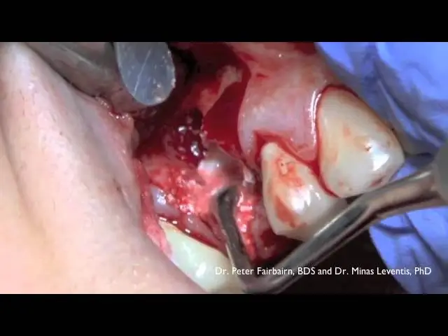
Often a year or more passed before an implant could support a new tooth. In the new protocol, the extraction socket is grafted soon after tooth extraction and implant placement can occur approximately 3 months after tooth extraction resulting in a significantly shorter treatment time. The product was developed in the UK and has been used in Europe successfully for 14 years. Please click on following video to see the product in use.
To be accepted for the study, patients must be at least 18 years of age and have no history of alcohol abuse, major health issues such as diabetes, autoimmune disorders, current or history of cancer, radiotherapy of head or neck. Also, patients cannot smoke or be currently pregnant.
Pre and post-surgical evaluation included CBCT evaluation will be taken without charge to the patient. In compensation for participation, all patients will receive a substantial discount off our standard fees. Patients who are accepted in the program must sign a release that will allow images of their tooth to be published in a scientific journal.
No facial photos or patient information will be included in the publication.

Histomorphometry of EthOss produced 51% new bone at 10 weeks
Fairbairn P, Leventis M, Van Stralen K, Horowitz R
Presented at ATTOI Tel Aviv May 22-24, 2017
If you would like to be considered for inclusion in this study, please call Kenneth M. Van Stralen, DDS office Phone Number 703-317-3900 to learn the details.
Conventional bone grafting techniques
We routinely perform grafting techniques using a combination of human and bovine source bone for socket grafting after tooth loss, ridge augmentation and sinus grafting prior to or in conjunction with implant placement. At times, we use a small amount of your own bone in severely atrophy cases, however, the majority of our grafting cases require no foreign substances or secondary surgical sites.
Severely atrophic maxilla’s (upper jaw) will likely require sinus grafting, however, we have found that in many cases, we can use bone from the implant procedure itself to gain the additional bone volume needed to complete the case. A limited (or internal) sinus graft also works well to a traumatically develop bone in the posterior maxilla.
Sinus Lift Procedure
The maxillary sinuses are above your of the upper back teeth. Sinuses warm and filter air. A healthy sinus is a hollow cavity. Some of the roots of the natural upper teeth extend up into the maxillary sinuses.
When these upper teeth are removed, the sinus will expand to leave a thin wall of bone separating the maxillary sinus from the inside of your mouth. If sufficient bone remains immediately after tooth extraction, the bone can be added to by grafting to allow placement of dental implants to keep the sinuses from expanding.
If the socket is not grafted immediately after extraction and the sinuses are allowed to expand, the floor of the sinus becomes very thin, it is impossible to place dental implants in this bone without a sinus graft.
A sinus graft or sinus lift is performed by entering the sinus from the side near where the upper teeth roots once were. A membrane lines the floor of the sinus. It is gently lifted upward and bone is inserted onto the floor of the sinus(or roof of the upper jaw). After several months of healing, the bone becomes part of the patient’s jaw and dental implants can be inserted and stabilized in this new sinus bone.
With sinus grafting,it possible for many patients to have dental implants and fixed restorations.Years ago there was no option for patients who had loose ill-fitting upper dentures.
If enough bone exists between the inside of your mouth and the bottom of the sinus is available to stabilize the implant, sinus augmentations and implant placement can be performed as a single procedure. If not enough bone is available, the sinus augmentation is performed first and the graft is allowed to mature for several months. Once the graft has matured, the implants can be placed.
Ridge Expansion
In severe cases of bone atropy, the bony ridge has been resorbed to point where insufficient bone width exist to place an implant that is covered in bone. One technique to increase ridge width is a ridge expansion.
In this procedure, the bony ridge of the jaw is gently separated by a series of ever larger screw. Bone graft material is placed in the newly opened space and allowed mature for a few months before placing the implant. In some instances, the implant can be placed at the same time the ridge is expanded.
Learn more about bone grafting and schedule your appointment today!